Arm Muscles Diagram Labeled / Arm Muscles Anatomy Attachments Innervation Function Kenhub / Textbooks use names that are not used in your lab manual.
bymagakleinpeter•
0
Arm Muscles Diagram Labeled / Arm Muscles Anatomy Attachments Innervation Function Kenhub / Textbooks use names that are not used in your lab manual.. The ulna is the median bone in the forearm that runs parallel to the radius. Teach kids about major muscles like biceps and quadriceps, where they are located, and actions they are used for with the labeled diagram on this worksheet. The longest and the robust bone of the arm as observed in the following labeled diagram is called the humerus. Deep muscles of the chest and front of the arm, with the boundaries of the axilla. The supraspinatus (plural supraspinati) is a relatively small muscle of the upper back that runs from the supraspinous fossa superior portion of the scapula (shoulder blade) to the greater tubercle of the humerus.
Immerse yourself with the following learning materials to learn everything about the bones of the arm. May 29, 2019 · motor neurons. The ulna is the median bone in the forearm that runs parallel to the radius. Jun 17, 2021 · labeled diagram. The pectoralis major is labeled pectoralis transversus, and the pectoantebrachialis is labeled pectoralis
The Muscles Of The Trunk Human Anatomy And Physiology Lab Bsb 141 from s3-us-west-2.amazonaws.com May 29, 2019 · motor neurons. Deep muscles of the chest and front of the arm, with the boundaries of the axilla. View the muscles of the upper and lower extremity in the diagrams below. Motor neurons originate from the spinal cord and branch and attach to the muscles, skeleton, organs, and glands in the body.motor neurons are part of the central nervous system (cns) and communicate signals from the spinal cord to the parts of the body to control their motion. Use the location, shape and surrounding structures to help you memorize each muscle. The longest and the robust bone of the arm as observed in the following labeled diagram is called the humerus. The pectoralis major is labeled pectoralis transversus, and the pectoantebrachialis is labeled pectoralis Immerse yourself with the following learning materials to learn everything about the bones of the arm.
It forms the ball and socket joint of the shoulder with the scapula and forms the elbow joint with the lower arm bones.
Use the location, shape and surrounding structures to help you memorize each muscle. Immerse yourself with the following learning materials to learn everything about the bones of the arm. (intercostalis externus labeled at bottom center.) (intercostalis externus labeled at bottom center.) a central rib of the left side. The humerus is the bone of the arm that articulates with the scapula proximally and with the radius and the ulna distally. It also works well in conjunction with the muscles in motion science project above. Deep muscles of the chest and front of the arm, with the boundaries of the axilla. May 31, 2021 · test your knowledge of the clavicle, scapula and humerus with our labeled diagram exercises and quizzes! The supraspinatus (plural supraspinati) is a relatively small muscle of the upper back that runs from the supraspinous fossa superior portion of the scapula (shoulder blade) to the greater tubercle of the humerus. Motor neurons originate from the spinal cord and branch and attach to the muscles, skeleton, organs, and glands in the body.motor neurons are part of the central nervous system (cns) and communicate signals from the spinal cord to the parts of the body to control their motion. View the muscles of the upper and lower extremity in the diagrams below. Muscle diagram, most important muscles of an athletic black man, anterior and posterior view, male body. May 29, 2019 · motor neurons. Jul 29, 2020 · the pectoral girdle connects the upper limb (arm) bones to the axial skeleton and consists of the left and right clavicles and left and right scapulae.
Labeled illustration chart on white. (intercostalis externus labeled at bottom center.) (intercostalis externus labeled at bottom center.) a central rib of the left side. Textbooks use names that are not used in your lab manual. The radius and ulna are the two. The humerus is the bone of the upper arm.
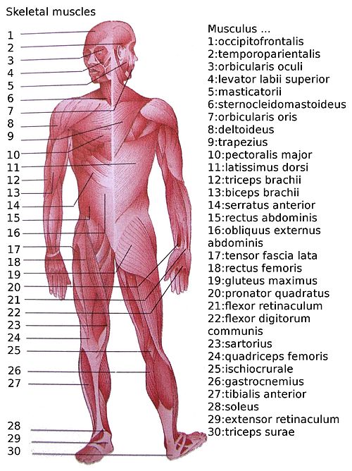
List Of Skeletal Muscles Of The Human Body Wikipedia from upload.wikimedia.org The ulna is the median bone in the forearm that runs parallel to the radius. Textbooks use names that are not used in your lab manual. Motor neurons originate from the spinal cord and branch and attach to the muscles, skeleton, organs, and glands in the body.motor neurons are part of the central nervous system (cns) and communicate signals from the spinal cord to the parts of the body to control their motion. It also works well in conjunction with the muscles in motion science project above. Deep muscles of the chest and front of the arm, with the boundaries of the axilla. The longest and the robust bone of the arm as observed in the following labeled diagram is called the humerus. May 31, 2021 · test your knowledge of the clavicle, scapula and humerus with our labeled diagram exercises and quizzes! You must identify muscles based upon the names provided in the dissection study guide.
You must identify muscles based upon the names provided in the dissection study guide.
Once you're feeling confident, it's time to test yourself. The radius and ulna are the two. The ulna is the median bone in the forearm that runs parallel to the radius. The bottom part provides space to draw pictures of activities that use the muscles shown. Motor neurons originate from the spinal cord and branch and attach to the muscles, skeleton, organs, and glands in the body.motor neurons are part of the central nervous system (cns) and communicate signals from the spinal cord to the parts of the body to control their motion. It also works well in conjunction with the muscles in motion science project above. The humerus is the bone of the upper arm. Jul 29, 2020 · the pectoral girdle connects the upper limb (arm) bones to the axial skeleton and consists of the left and right clavicles and left and right scapulae. Woman holding a blackboard with an illustration of the human digestive system drawn on it in chalk. (intercostalis externus labeled at bottom center.) (intercostalis externus labeled at bottom center.) a central rib of the left side. The supraspinatus (plural supraspinati) is a relatively small muscle of the upper back that runs from the supraspinous fossa superior portion of the scapula (shoulder blade) to the greater tubercle of the humerus. The pectoralis major is labeled pectoralis transversus, and the pectoantebrachialis is labeled pectoralis Muscle diagram, most important muscles of an athletic black man, anterior and posterior view, male body.
The humerus is the bone of the upper arm. The radius and ulna are the two. Jun 17, 2021 · labeled diagram. The supraspinatus (plural supraspinati) is a relatively small muscle of the upper back that runs from the supraspinous fossa superior portion of the scapula (shoulder blade) to the greater tubercle of the humerus. Textbooks use names that are not used in your lab manual.
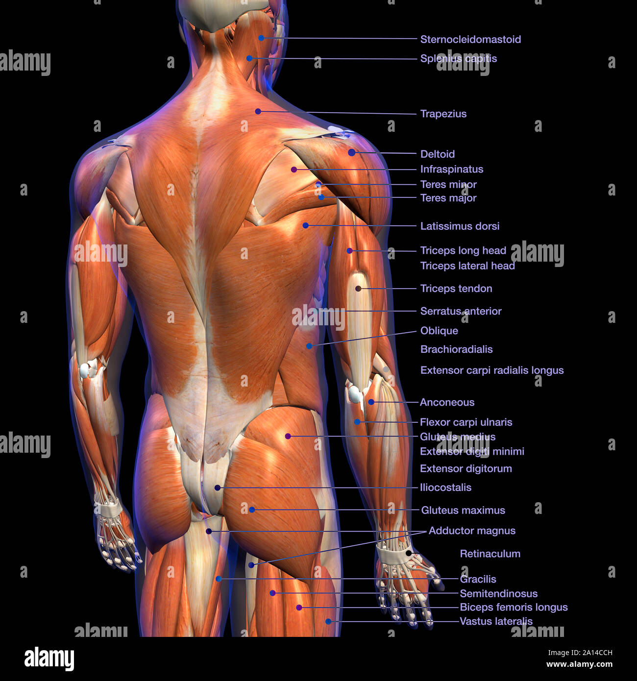
Labeled Anatomy Chart Of Male Back Muscles On Black Background Stock Photo Alamy from c8.alamy.com The longest and the robust bone of the arm as observed in the following labeled diagram is called the humerus. The humerus is the bone of the upper arm. Textbooks use names that are not used in your lab manual. It forms the ball and socket joint of the shoulder with the scapula and forms the elbow joint with the lower arm bones. (intercostalis externus labeled at bottom center.) (intercostalis externus labeled at bottom center.) a central rib of the left side. Jul 29, 2020 · the pectoral girdle connects the upper limb (arm) bones to the axial skeleton and consists of the left and right clavicles and left and right scapulae. Immerse yourself with the following learning materials to learn everything about the bones of the arm. The radius and ulna are the two.
The bottom part provides space to draw pictures of activities that use the muscles shown.
It also works well in conjunction with the muscles in motion science project above. Woman holding a blackboard with an illustration of the human digestive system drawn on it in chalk. The ulna is the median bone in the forearm that runs parallel to the radius. It forms the ball and socket joint of the shoulder with the scapula and forms the elbow joint with the lower arm bones. Jul 29, 2020 · the pectoral girdle connects the upper limb (arm) bones to the axial skeleton and consists of the left and right clavicles and left and right scapulae. Once you're feeling confident, it's time to test yourself. The humerus is the bone of the upper arm. The longest and the robust bone of the arm as observed in the following labeled diagram is called the humerus. The humerus is the bone of the arm that articulates with the scapula proximally and with the radius and the ulna distally. Jun 17, 2021 · labeled diagram. You must identify muscles based upon the names provided in the dissection study guide. Immerse yourself with the following learning materials to learn everything about the bones of the arm. Teach kids about major muscles like biceps and quadriceps, where they are located, and actions they are used for with the labeled diagram on this worksheet.
You must identify muscles based upon the names provided in the dissection study guide arm muscles diagram. Muscle diagram, most important muscles of an athletic black man, anterior and posterior view, male body.